SPECT: Imaging Blood Flow and Function in the Body
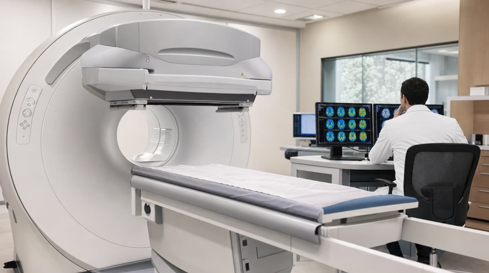
SPECT stands for Single Photon Emission Computed Tomography. It is a nuclear imaging test that produces three-dimensional pictures of how blood flows to tissues and organs — in other words, it shows function, not just structure. That distinction is what makes it uniquely useful for studying the brain, the heart, and any condition where the problem is reduced blood supply, or hypoxia.
How a SPECT scan works
Before the scan, a small amount of a radioactive tracer (a “radiopharmaceutical,” often technetium-99m) is injected into a vein. The tracer travels in the bloodstream and accumulates in tissues in proportion to how much blood they receive. Areas with healthy blood flow take up more tracer; areas that are under-perfused take up less.
The tracer emits gamma rays — single photons — as it decays. A rotating gamma camera detects those photons from many angles around the body, and a computer reconstructs the signals into cross-sectional slices, much as a CT scanner builds an image from X-rays. The result is a color-coded map where brightness corresponds to blood flow or metabolic activity.
What SPECT is used for
Because it images function, SPECT answers questions that anatomical scans cannot:
- Brain perfusion. Mapping regional cerebral blood flow in stroke, traumatic brain injury, and dementia — showing which regions are metabolically quiet even when they look structurally intact.
- Cardiac (myocardial) perfusion. Revealing areas of heart muscle that receive too little blood during stress, a mainstay of coronary artery disease assessment.
- Bone and infection imaging. Locating stress fractures, infection, or bone necrosis that plain X-rays miss.
SPECT vs CT, MRI and PET
These tests are often confused, so it helps to line them up:
- CT uses X-rays to show detailed anatomy — bones, bleeds, tumors.
- MRI uses magnetic fields to show soft-tissue anatomyin high detail, without radiation.
- PET (positron emission tomography) also images function and metabolism, generally at higher resolution than SPECT but at greater cost.
- SPECT images function and blood flow using widely available, longer-lived tracers — making it more accessible than PET for perfusion studies.
In practice the tests are complementary: a CT or MRI shows what a region looks like, while a SPECT shows whether it is working.
Why SPECT matters in oxygen and hyperbaric medicine
Many conditions treated with hyperbaric oxygen therapy involve tissue that is alive but starved of oxygen — the “ischemic penumbra” around an injury. Because SPECT maps blood flow directly, it has been used in research to visualize regional cerebral perfusion in people with brain hypoxia and brain injury, and to document changes in that perfusion. This is precisely why brain-perfusion SPECT appears so often in discussions of HBOT for neurological conditions.
A word of caution: imaging that a region’s blood flow changed is not the same as proving a treatment made someone better. SPECT is a powerful window into function, but its findings are interpreted alongside symptoms, clinical exams and controlled research — never in isolation.
Is a SPECT scan safe?
SPECT is generally considered safe. It does involve a small dose of ionizing radiation from the tracer, comparable to other common nuclear medicine tests, and the tracer clears from the body over hours to a day or two. As with any imaging that uses radiation, the benefits and risks are weighed by the physician ordering the scan, and it is generally avoided in pregnancy unless essential.
SPECT perfusion maps are often used to document brain blood-flow changes after a course of pressurized oxygen. For how those treatment sessions are actually delivered, see this overview of modern HBOT protocols.
For detailed medical information, please consult with a qualified healthcare professional.
Sources
How oxygen shortage affects the brain — the function SPECT helps visualize.
Hyperbaric oxygen therapy indications, protocols and evidence.
The oxygen-shortage states behind reduced perfusion on a scan.