Thermal Burns

Thermal Burns can be caused to the skin by fire, flame, steam, hot liquids or one of several heating devices. Other sources of burns are those cased by chemicals, radiation, sun, or electricity. By severity the thermal burn injury can be classified as first, second or third degree. Burns severity, extent as well as the number of affected skin layers determine symptoms experienced by a victim.
©2008 Undersea and Hyperbaric Medical Society, Inc. The book can be purchased at UHMS web site.
By Paul Cianci, M.D. and J. Benjamin Slade, Jr., M.D.
Severe thermal injury is one of the most devastating physical and psychological injuries a person can suffer. In the United States, more than 2 million burn injuries require medical attention each year, resulting in 14,000 deaths. About 75,000 patients require hospitalization each year, and 25,000 of those remain hospitalized for more than 2 months. The most common mechanisms of burn injury are flame and scalding, and the upper extremity, head and neck are the most common body areas involved.
The average cost of care for a critically burned patient in 1992 ranged from $23,000 to $34,000. Adjusting for 10% inflation/year, current average cost would range from $60,000 to $88,000. According to the executive director of the American Burn Association, for patients who survive 60% total body surface area burns (TBSA), charges for the hospital stay alone, not including operating room time, surgeons bill, artificial skin, rehabilitation, and other costs, averages $400,000 (in 2003 U.S. dollars).
These costs can reach $500,000 for burns over 80% TBSA. During 1997-98, in a northern California regional burn center, hospital costs for 20 burn patients averaged $253,000 (range $1,100 to $1.5 million) per patient. This includes the cost of hyperbaric oxygen (HBO2) which averaged $6,360 (range $1,000 to $27,500) per patient.
The burn wound is a complex, dynamic injury characterized by a zone of coagulation (or complete capillary occlusion), surrounded by an area of stasis, and bordered by an area of erythema. The zone of coagulation may increase by a factor of 10 during the first 48 hours after injury. Ischemic necrosis quickly follows. Hematological changes, including platelet microthrombi and hemoconcentration, occur in the postcapillary venules. Edema formation is rapid in the area of injury but also develops in distant, uninjured tissue.
There are concurrent changes occurring in the distant microvasculature where red cell aggregation, white cell adhesion to venular walls, and platelet thromboemboli occur. Inflammatory mediators are elaborated locally in part from activated platelets, macrophages, and leukocytes, contribute to localand systemic hyperpermeability of the microcirculation and appear histologically as gaps in the venular and capillary endothelium. This progressive process may extend damage dramatically during the early days after injury.
The continuing tissue damage in thermal injury is due to the failure of the surrounding tissue to supply borderline cells with oxygen and nutrients necessary to sustain viability. The impediment of circulation below the injury leads to desiccation, as fluid cannot be supplied via the thrombosed or obstructed capillaries. Topical agents and dressings may reduce but do not prevent desiccation of the burn wound and the inexorable progression of injury to deeper layers.
Infection remains the leading overall cause of death from thermal burns. Susceptibility to infection is greatly increased due to the loss of the integumentary barrier to bacterial invasion, the ideal substrate present in the burn wound, and the compromised or obstructed microvasculature which prevents humoral and cellular elements from reaching the injured tissue.
Additionally, the immune system is seriously affected, demonstrating decreased levels of immunoglobulins and serious perturbations of polymorphonuclear leukocyte (PMNL) function including a reduction in chemotaxis, phagocytosis, and diminished killing ability, resulting in increased morbidity and mortality. Regeneration cannot take place until equilibrium is reached; hence, healing is retarded. Prolongation of the healing process may lead to excessive scarring.
Hypertrophic scars are seen in only 4% of cases requiring 10 days to heal, but up to 40% of cases requiring longer than 21 days to heal. Therapy of burns, therefore, must by directed to minimizing edema, preserving marginally viable tissue, enhancement of host defenses, and promoting wound closure.
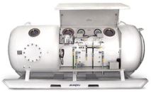
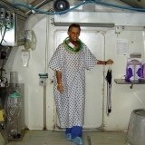
Adjunctive Hyperbaric Oxygen Therapy can benefit each of these problems directly, and shows promise in the treatment of inhalation injury. The more extensive the thermal burns injury, the higher the incidence of an inhalation injury and pulmonary injury caused by smoke inhalation is the primary cause of fire-related deaths. The airway injury can be worsened by a variety of chemical pyrolysis products, depending on the material burned.121
Summary
Current data show that Hyperbaric Oxygen Therapy, when used as an adjunct in a comprehensive program of thermal burns care, can significantly improve morbidity and mortality, reduce length of hospital stay, and lesson the need for surgery. It has been demonstrated to be safe in the hands of those thoroughly trained in rendering Hyperbaric Oxygen Therapy in the critical care setting and with appropriate monitoring precautions. Careful patient selection and screening is mandatory.