Retinopathy Of Prematurity.
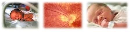
Retinopathy of prematurity (ROP) is a potentially blinding eye disorder that mainly affects premature born infants. The article published below highlights the tragic failure of mainstream medicine to acknowledge the use of oxygen under pressure in treatment of retinopathy of prematurity. Prof. Philip James, M.D. of Dundee University, Scotland insists that it is withdrawal of oxygen that causes blindness of newborn babies. This has resulted in millions of infants not being properly treated.
Featured article.
Retinopathy Of The Newborn Is Due To Hypoxia Not Oxygen Toxicity.
Philip B James MB ChB, PhD, DIH, FFOM. Wolfson Hyperbaric Medicine Unit, Ninewells Hospital and Medical School, Dundee, Scotland UK.
Summary
The use of oxygen in incubators for the newborn has been restricted to a maximum level of 40% based on the recorded epidemics of blindness in the 1950's. Prevailing opinion attributes the retinopathy to oxygen toxicity, but the evidence indicates that it is due to the suppression of the development of the retinal microcirculation and the hypoxia that results from the subsequent lowering of the level of oxygen. Alternative strategies, using intermittent high dosages of oxygen may avoid the retinopathy and by improving the outcome for the brain thereby reduce the incidence of cerebral palsy.
Introduction
The introduction of oxygen tents and incubators following World War II allowed premature infants to be given supplementary oxygen to improve their chances of survival and levels up to 70% were given for extended periods. Epidemics of blindness followed in the 1950's1 and led to a restriction of the level of supplemental oxygen to 40%. This was followed by a dramatic reduction of the incidence of the retinopathy which confirmed the involvement of oxygen. Since then every medical student has been taught that the retinopathy of the premature (ROP) is caused by oxygen toxicity.
However, in 1992 a Lancet editorial2 observed that even the use of continuous transcutaneous monitoring to avoid transient hyperoxia has failed to abolish the retinopathy and discussed the adverse effect of oxygen restriction on mortality. Despite many improvements in neonatal care the problem of cerebral palsy remains and McDonald3 recorded a significant rise in the incidence of cerebral palsy with the restriction of oxygen levels in a comparison of rates before and after the restriction.
Materials And Methods
A survey of the literature has been undertaken to examine both the clinical and experimental observations relating to retrolental fibroplasia. Szewczyk4 was the first to suggest that retinopathy of the premature was produced by too rapid a reduction of the level of oxygen after a child had been habituated to an enriched oxygen atmosphere. He found the condition could actually be treated by returning the infant to the high oxygen level, followed by a slow return to breathing air. A small controlled trial was undertaken with 24 premature infants being slowly reduced to a normal atmospheric concentration and 26 abruptly withdrawn.5 Only 2 infants in the first group developed retinopathy compared to 13 in the second. (p<0.001)
However the study was criticized because the infants who were in the weaned group actually had less oxygen. Nevertheless it is clear that the investigators only undertook the study because they knew that the retinopathy could be reversed by re-exposing the infants to a raised partial pressure of oxygen. A large multicentre study6 was finally undertaken which appeared to settle the issue, but although the link to the use of oxygen was certainly proven, it did not establish that the retinopathy is caused by the toxicity of oxygen. There are a number of inconsistencies in the data which are linked to spontaneous regression. Forrester7 also returned infants who had developed a retinopathy to a high level of oxygen.
In 1964 he reported a study of 17 such children stating "the results were spectacular; in each individual case the retinal vascular pattern, having shown gross abnormalities, returned to normal." In most cases return to a high concentration and a slow reduction of the level of oxygen to an atmospheric concentration over a period of weeks was all that was necessary to solve the problem. Two infants needed a third period of oxygen exposure, because the disease again became active.
Of the seventeen infants twelve recovered with normal eyes and five had minor permanent changes not causing blindness. Many of these infants were therefore exposed to high oxygen tensions for very long periods. (the longest were 93, 88, 85 and 83 days) He commented; "if one believes, that oxygen has a direct toxic effect on the infant's retina these surely would have been the infants who became blind, for they were all of very low birth weight, all had the early retinopathy and they were all subjected to intensive and prolonged therapy." Others made the same observations 8,9,10,11 but examination of the withdrawal periods used by other researchers indicates that none used the extended durations used by Forrester.
The key question is:- do the changes develop during the time at the high partial pressure? The sequence of the vascular changes suggests that the retinopathy is due to hypoxia and it is relevant that the only established risk factors in retinopathy of the premature are conditions associated with intrauterine hypoxia.12 Initially the vessels dilate, becoming tortuous and this is followed by oedema and haemorrhage. Vascular dilatation and increased permeability occur in response to hypoxia and diapedetic haemorrhage indicates gross impairment of the blood-retinal barrier.
The developing oedema will begin to separate the retina and also impair oxygen transport. Because the retinal vessels are applied to the concave hemisphere of the globe of the eye it is reasonable to suggest that the tortuosity occurs because the scleral tissues are inelastic and mechanical distortion contributes to the retinal detachment. If this is the case then increasing the plasma oxygen tension, which can paradoxically induce vasoconstriction whilst increasing the gradient for oxygen transport, would be expected to reverse the condition as described.
Recently, hypoxia has been implicated in the glial reaction in and experimental model of retinal detachment.13 Lewis et a114 have studied the effect of oxygen supplementation in this model. They found that oxygen supplementation at 70% limited the proliferation of the retinal Muller cells responsible for gliosis and assisted in maintaining their normal structure and function. It is notable that the percentage of oxygen used was typical of the values implicated in ROP.
The level of oxygen determines the capillary density in a tissue even in the adult mammalian brain. Rats decompressed to half atmospheric pressure for three weeks increase the capillary density in the brain by 50%.14 This mechanism is part of the adaptation to altitude that allows climbers to ascend to very high altitudes, such as Mount Everest, without supplementary oxygen. Hyperoxia has the opposite effect in the developing retina.
Ashton et al15 is demonstrated that oxygen supplementation suppressed the development of the retinal circulation in an experimental model. As the choroidal circulation and the vitreous provide oxygen to support the retina, this could perhaps be expected. The circulation of both the brain and the retina mature rapidly in the last few weeks of pregnancy16 and the maintenance of hyperoxia would predictably suppress vessel formation, not because of oxygen toxicity, but because of the absence of the stimulus of hypoxia.
Results And Conclusions
The data confirm that the modulation of microcirculatory development by long-term exposure to high levels of oxygen is responsible for the later development of retinopathy. This occurs because the vasculature is not able to supply sufficient oxygen when the infant finally breathes air. The intermittent use of higher dosages of oxygen may allow the normal development of the retinal circulation and yet prevent the retinopathy of prematurity. Oxygen as a cerebro-protective agent may also provide the much sought after intervention to lower the incidence of cerebral palsy.
References
1. Forrester RM, Jefferson E, Naunton WJ. Oxygen and retrolental fibroplasia; a seven-year survey. Lancet 1954;ii:258-260.
2. Editorial. Oxygen restriction and retinopathy of prematurity. Lancet 1992;339:961-962.
3. McDonald AD. Oxygen Treatment of Premature Babies and Cerebral Palsy. Dev Med Child Neurol 1964;6:313-314.
4. Szewczyk TS. Retrolental fibroplasia: etiology and prophylaxis. Amer J Ophthalmol;1951:34:1609.
5. Bedrossian RH, Carmichael P, Ritter J. Retinopathy of prematurity (retrolental fibroplasia) and oxygen. Amer J Ophthalmol 1954;37:78.
6. Kinsey VE. Retrolental fibroplasia. Co-operative study of retrolental fibroplasia and the use of oxygen. Arch Ophthalmol 1955;59:481-542.
7. Forrester RM. Oxygen cerebral palsy and retrolental fibroplasia. Dev Med Child Neurol 1964;6:648-650.
8. Swart-Van Der Hoeven JT, Mak, TMB. Effects of oxygen on retrolental fibroplasia in premature infants, with report of two cases. Maadschr Kindergeneesk 1952;20:276.
9. Von Winning, CHOM. Retrolental fibroplasia and other forms of pseudoglioma. The Hague Drukkerij Trio.
10. Tiddens H: A case of retrolental fibroplasia treated and cured with administration of oxygen in alternating concentrations. Ophthalmologica 1960;139:475-88.
11. Tanabe Y. Intermittent oxygen for the treatment of the retinopathy of prematurity. Nippon Ganka Gakkai Zasshi 1972 May 76:5 316-21.
12. Johnson I, Schaffer DB, Blessa MI. Factors predisposing to RLF: complications of pregnancy. Pediat Res 1980;14:601.
13. Lewis G, Mervin K, Valter K, Maslim J, et al. Limiting the proliferation and reactivity of retinal Muller cells during experimental retinal detachment: the value of oxygen suplementation. Am J Ophthalmol 1999;128:165-172.
14. Hank SI, Behmand RA, LaManna JC. Hypoxia increases glucose transport at blood-brain barrier in rats. J Appl Physiol 1994;77:896-901.
15. Ashton N, Garner A, Knight G. Intermittent oxygen in retrolental fibroplasia. Am J Ophthalmol 1971;71:153-160.
16. Takashima S, Tanaka K. Development of cerebrovascular architecture and its relationship to periventricular leukomalacia. Arch Neurol 1979;35:11-16.
For PDF version of this article click here...
Patients with Retinopathy Of Prematurity are at greater risk for strabismus, glaucoma, cataracts and myopia later in life, and should be examined yearly to help prevent and treat these conditions.
Sponsored links: