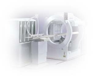
SPECT - Single Photon Emission Computed Tomography
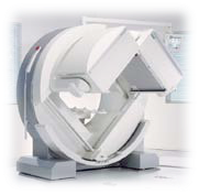
SPECT stands for Single Photon Emission Computed Tomography. It is a type of nuclear imaging test that shows how blood flows to tissues of organs.
This type of imaging shows brain function, as opposed to MRI or CT which show structure of organs such as a brain.
Tests have shown that Single Photon Emission Computed Tomography might be more sensitive to brain injury than either MRI or CT because it can detect reduced blood flow to injured sites.
Single Photon Emission Computed Tomography is also useful in diagnosing stress fractures in the spine (spondylolysis), blood deprived (ischemic) areas of brain following strokes and tumors.
In classical neurologic conditions such as stroke, brain trauma, Alzheimers or epilepsy / seizures Single Photon Emission Computed Tomography imaging has been often a lifesaving test. Traumatic Brain Injuries (TBI) as well as more subtle brain abnormalities previously undetected has been often functionally defined by Single Photon Emission Computed Tomography.
Single Photon Emission Computed Tomography imaging involves an intravenous injection of two substances a tracer (radioisotope) and a blood flow agent. The radioisotopes usually used in SPECT scan are iodine-123, technetium-99m, xenon-133, thallium-201, and fluorine-18. These radioactive forms of natural elements pass safely through the body and are detected by the scanner.
Various drugs and other chemicals can be labeled with these isotopes without changing their properties. The type of tracer used depends on what condition a physician wants to verify. For example, if a doctor is looking at a tumor, she might use radiolabeled glucose (FDG) and watch how it is metabolized by the tumor.
Utilization of SPECT is not restricted to brain imaging only. The following video demonstrates how SPECT technology is used in heart imaging...
Technetium is a low energy, radioactive element that is used in nearly all nuclear medicine scans in infants, children and adults. Ceretec and Neurolite are the names of the blood flow agents which bind to Technetium and when injected, distribute throughout the brain in proportion to blood flow. Brain scanning reveals images of blood flow at the time of injection. The total body radiation dose from injected Technetium is very small, being less than that which the chest or brain receives with an x-ray or CT scan. There are no reported side effects from injection of Ceretec or Neurolite.
Some behavioral-cognitive problems such as violent behavior, hyperactivity, compulsions, hyperactivity, poor attention, major depression, bipolar disorders, and anxiety disorders can be traced and analyzed by Single Photon Emission Computed Tomography.
Photon Emission Computed Tomography - How The Scan Is Performed...
First patient receives an injection of a small amount of radioactive tracer and asked to rest for about 1020 minutes until the tracer reaches the brain. Next she lies comfortably on a scanner table while a special camera rotates around patients head. Patient has to remain as still as possible so that the machine can get accurate pictures. Once the Single Photon Emission Computed Tomography scan is complete, patient can leave. Drinking plenty of water is essential after the test to flush out any tracer left in your body.
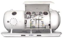
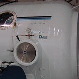
Photon Emission Computed Tomography and Hyperbaric Oxygen Therapy (HBOT)
The most recent and most significant documented advances with Hyperbaric Oxygen Treatment have emerged with the utilization of high tech investigation including Single Photon Emission Computed Tomography and isotopic tracers with Magnetic Resonance Imaging (MRI). SPECT performed as a pre and post hyperbaric evaluation provides valuable insights into the mechanisms and actions of Hyperbaric Oxygen Therapy through oxygenation.
The images below show SPECT scans of 17 years old male suffered from chronic neurologic deficiency and admitted to Hyperbaric Oxygen Treatment 1 month after moderate Traumatic Brain Injury (TBI). Status on admission to HBOT:
- Lt. Hemiparesis
- Severe coordintion disfunction
- Severe cognitive disfunction
- Normal Brain CT scan
SPECT imaging shows dramatic improvement of the chronic neurologic deficiency after 30 treatments with Hyperbaric Oxygen.
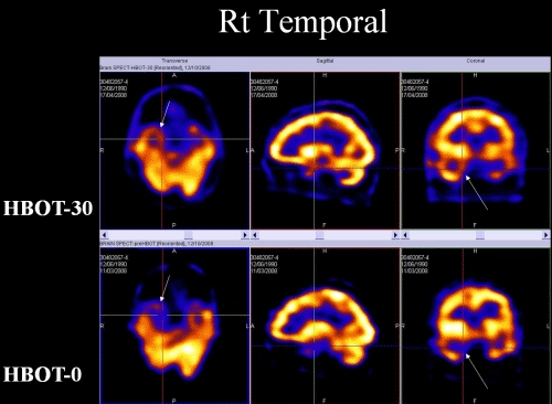
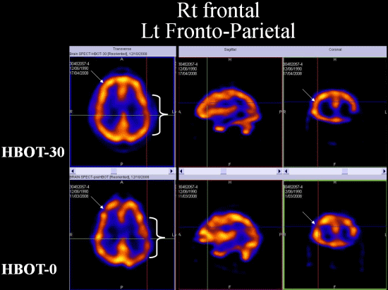
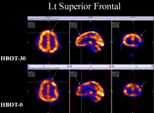
The above images was kindly provided by Dr. S. Efrati M.D., Head of the Institute of Hyperbaric Medicine, Head of Research & Development unit, Assaf-Harofeh Medical Center, Israel.
Conditions that have previously been considered to have a poor prognosis, including brain injuries, stroke and neurological based conditions, have been greatly improved with Hyperbaric Oxygen Therapy supported by Single Photon Emission Computed Tomography imaging and continue, today, to be among the areas of research.