Respiratory System Diagram

This respiratory system diagram presents a structure of the respiratory system as well as an external respiration process, or breathing.
The life actually occurs at a cellular level with Oxygen playing one of the most essential roles in biochemical and physiologic processes involved in cellular respiration.
The internal or cellular respiration process in which glucose or other small molecules are oxidized in order to produce the vital energy is required for survival and reproduction of our cells and tissues. The respiratory system comprises the air passages organs (described in this respiratory system diagram) and the diaphragm.
Breathing
Breathing is an external respiration process of extracting Oxygen from the air and returning back Carbon Dioxide which is an output product of a cellular respiration. The air we breathe is a mixture of gases consisting approximately of 21% Oxygen, 78% Nitrogen and 1% Carbon Dioxide or other gases and water vapors. The respiration process changes these proportions to approximately 16% of Oxygen, 78% of Nitrogen, 5% of Carbon Dioxide and 1% of other gases while water vapors saturation reaches 100%.
A simplified frontal section of respiratory system diagram showing the principal organs of the respiratory system.
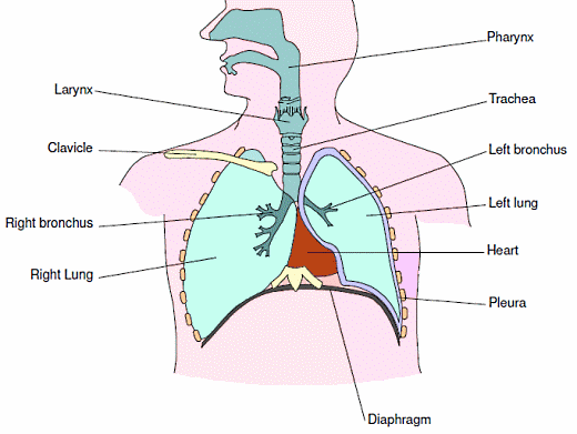
Respiratory System Anatomy
Nose
The nose is an irregular cavity at the anterior part of the face, with an external protuberance. The nose is divided in two by the nasal septum which is composed of cartilage. The floor of the nasal cavity is formed by the roof of the mouth or hard palate. The nasal cavity is lined with ciliated mucous membrane.
Respiratory System Diagram Of Nasal Cavity
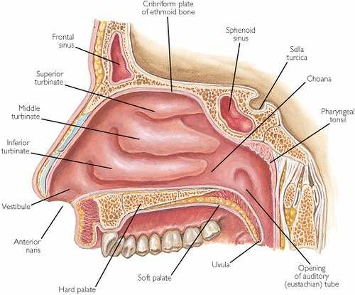
In the upper part of the nasal cavity, the endings of the olfactory nerve neurones provide the sense of smell. The irregular internal structure causes turbulence which spreads inhaled air over the surface where mucus absorbs dust and other contaminants, and provides a warming and moistening action. The nasal cavity opens into the pharynx
Pharynx
The pharynx is a cavity lying behind the nose and mouth. Inferiorly, the pharynx opens into the oesophagus and trachea. The pharynx is divided into three areas: the nasal pharynx, the oropharynx, and the laryngopharynx.
Respiratory System Diagram Of Pharynx
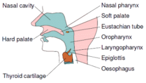
Nasal pharynx
The nasal pharynx is the area immediately superior to the soft palate. Like the nasal cavity, the nasal pharynx is lined with ciliated mucous membrane. Auditory tubes (Eustachian tubes) - one from each middle ear - open into the sides of the nasal pharynx.
Oropharynx
The oropharynx lies at the posterior part of the mouth, and extends from the soft palate to the epiglottis. A membranous passageway, known as the fauces, separates the oropharynx from the mouth. This passageway opens during swallowing or when inhaling through the mouth.
Laryngopharynx
The laryngopharynx is the lowest part of the pharynx and leads into the larynx. At this point the airway and digestive passages divide, the digestive passage continuing as the oesophagus.
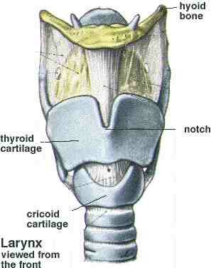
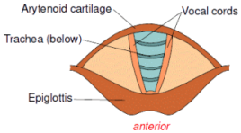
Larynx
The larynx forms the connection between the pharynx and the trachea. It also contains membranes (the vocal cords) which allow the production of sounds. The larynx is supported by the thyroid cartilage, the outline of which is visible as the "Adam's Apple". The opening into the larynx is protected by the epiglottis. This is a flap of fibrous tissue which moves over the larynx during swallowing so that solids and liquids are directed into the oesophagus.
Respiratory System Diagram Of Larynx
Vocal Cords
The vocal cords are made up from two strips of epithelium at the base of the larynx. The cords are supported between the thyroid cartilage and arytenoid cartilage. When muscles associated with the arytenoid cartilage contract, the vocal cords are pulled together, leaving only a small gap. In a process termed phonation, the vocal cords vibrate and produce sound as air is forced through the gap. This sound is then formed into the voice by movement of the tongue, mouth, and lips.
Respiratory System Diagram Of Vocal Cords
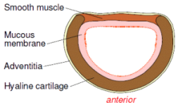
Trachea
The trachea is a tube which extends from the base of the larynx to the lungs, where it divides into two bronchi. The trachea is formed from connective tissue and smooth muscle, supported by 16 to 20 "C" shaped incomplete rings of cartilage. The cartilage prevents collapse of the trachea, while the open section of the "C" shape allows dilation and contraction under control of the nervous system, to enhance respiratory effort when necessary. The trachea is formed from three layers of tissue:
- Outer layer. This is composed of fibrous and elastic tissue.
- Middle layer. This is composed of cartilage and bands of smooth muscle.
- Inner layer. This is composed of ciliated mucous membrane.
Respiratory System Diagram Of Trachea
Bronchi and Bronchioles
The bronchi are two tubes which originate at the division of the trachea. The left bronchus is slightly longer than the right as it has to pass around the heart to reach the left lung.
Respiratory System Diagram Of Bronchi and Bronchioles
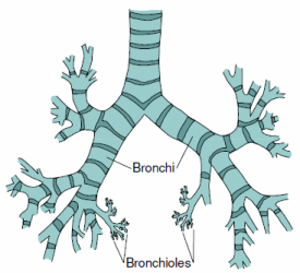
Each bronchus leads into a lung, entering together with the pulmonary blood vessels, at a point known as the hilum. Inside the lungs, the bronchi divide, and subdivide, into progressively smaller and more numerous, bronchioles. The bronchi have the same tissue formation as the trachea, allowing similar autonomic nervous control, although the amount of cartilage diminishes towards their distal ends, and is not present in bronchioles. The bronchioles end in very fine alveolar ducts leading to the alveoli.
Respiratory System Diagram Of Alveoli
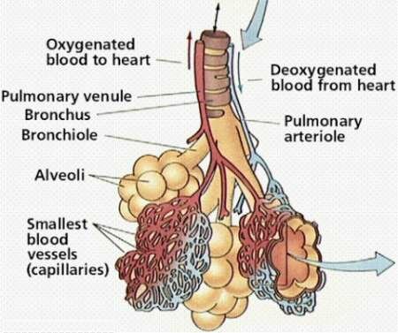
Alveoli
The alveoli, or air sacs, are the ends of the air passages. They are arranged in bunches, each bunch forming the end of a single bronchiole. Each alveolus is closely surrounded by blood capillaries so as to allow the exchange of gases to take place between the blood and the inhaled air.
The alveolar walls are composed of a single layer of endothelium - only about 4µm in thickness, allowing the passage of gas, and even individual red blood cells, to and from the surrounding capillaries. Each lung contains up to 350 million alveoli, giving a total gas exchange surface of approximately 65 m2.
Respiratory System Diagram Of Alveoli

Lungs
The lungs, being the combination of the bronchi, bronchioles, alveoli, and their connecting blood vessels, occupy the majority of the thorax. The area between the lungs, the mediastinum, is occupied by the heart and major vessels.
Respiratory System Diagram Of Lungs
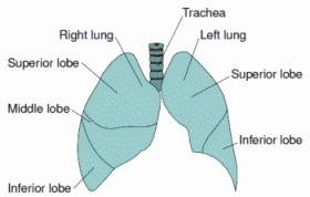
The lungs are subdivided into lobes - the right lung having three: the superior, the middle, the inferior - the smaller left lung having two: the superior, and the inferior. (the left lung is smaller as part of its potential volume is occupied by the heart). Each lobe is fed by a major division of the appropriate bronchus. You can’t breathe and swallow at the same time – this prevents you from choking! Every minute we breathe in about 13 pints of air making about 20 breaths each minute.
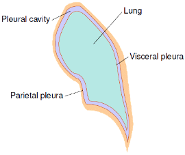
Pleurae
Each lung is enclosed in a membranous sac, or pleura. There are two layers of pleura: Parietal pleura. This is the outer membrane, which is attached to the inside of the chest wall, and the superior surface of the diaphragm.
Visceral pleura. This is the inner membrane, which is attached to the outer surface of the lung. The pleurae are composed of serous membrane which secretes fluid into the potential space, the pleural cavity, between the parietal and visceral layers. This fluid provides lubrication for the movement which takes place during respiration.
Respiratory System Diagram Of Pleurae
The diaphragm is the muscle that moves up or down to increase or decrease the chest cavity volume. Hiccups are muscle spasms of the diaphragm. Sneezing is like a cough in the upper breathing passages. It is a body’s way of removing an irritant from the sensitive mucus membranes.
You shouldn’t breathe through your mouth! Your nasal cavity is lined with cilia (tiny hairs) and mucus to filter out bacteria, allergens, fungus, and other irritants. When you breathe through your mouth, these things are not filtered out as efficiently which means you can get sick easier!
To visualize the respiratory system diagram please watch this video...
Return to the Hyperbaric Oxygen (HBO2) Overview